Figure 3.
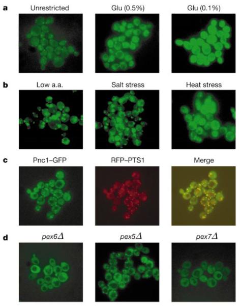
Pnc1–GFP is localized in the nucleus and cytoplasm, and concentrated in peroxisomes. a, Pnc1–GFP fluorescence in glucose-restricted cells (Glu 0.5% and 0.1%). b, Pnc1–GFP fluorescence under conditions of mild stress (a.a., amino acid). c, Co-localization of Pnc1–GFP (green) and RFP–PTS1 (red). Yellow indicates overlap. d, Localization of Pnc1–GFP in cells from peroxisomal mutant strains, pex6Δ, pex5Δ and pex7Δ.

