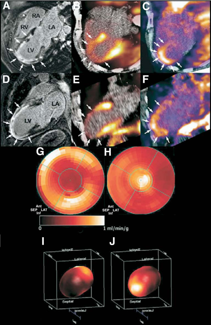
Figure 10. Clinical PET Imaging of Infarct Angiogenesis.
(A) Four- and (D) 2-chamber MR images after gadolinium administration reveal delayed enhancement (arrows) of the anterior wall and apical regions corresponding with (B, E) anatomically matched severely decreased myocardial perfusion (arrows) by 13N-ammonia radionuclide scanning. (C, F) Focal 18FDG-PET uptake colocalized (arrows) with the infarct zone, possibly representing regions of neovascularization. (G) Two- and (I) 3-dimensional polar maps of 13N-ammonia myocardial blood flow demonstrate a significant reduction in the distal left anterior descending artery territory. (H, J) The 18F-Galakto-RGD signal on PET-imaging coregistered with areas of severely diminished 13N-ammonia perfusion. Reproduced, with permission, from Makowski et al. (28). RGD = Arginine-Glycine-Aspartate peptide motif; 13N = nitrogen N 13; other abbreviations as in Figures 2, 7, and 8.

