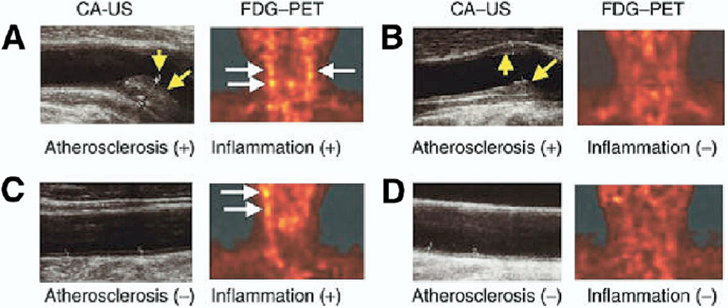
Figure 8. Clinical PET Imaging of Atherosclerosis Inflammation.
Paired carotid 18FGD-PET and B-mode ultrasound images of human patients having atherosclerotic disease (A) with or (B) without 18FDG-derived evidence of plaque inflammation and patients without detectable atherosclerosis (C) with or (D) without inflammation. White arrows = 18FDG-PET signal uptake. Yellow arrows = carotid atherosclerotic plaque. Reproduced, with permission, from Tahara et al. (36). CA-US = carotid atherosclerosis ultrasound; 18FGD = fluorodeoxygenase F 18; other abbreviations as in Figure 7.

