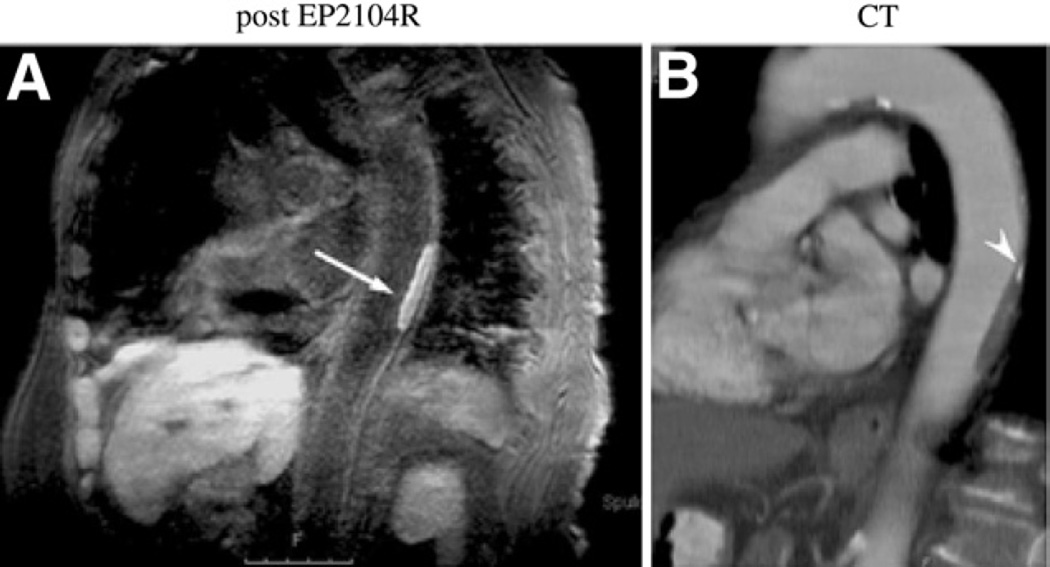
Figure 9. Clinical CMR of Fibrin-Rich Thrombi.
(A) Magnetic resonance molecular imaging of a descending aortic thrombus with a fibrin-specific probe (EP-2104R) in an 82-year-old female patient. The thrombus is visualized as positive contrast (arrow) on the inversion recovery black-blood gradient-echo imaging sequence. (B) Reconstructed images from a corresponding contrast-enhanced CT confirm the thrombotic filling defect and note a small region of calcification (arrow-head). Reproduced, with permission, from Spuentrup et al. (40). Abbreviations as in Figures 2 and 7.

