Fig. 5.

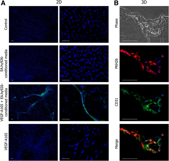
Endothelial differentiation of UC-MSCs. a Endothelial differentiation of UC-MSCs in monolayer. Three endothelial induction media were used for UC-MSC culture: EA.hy926-conditioned media mixed 1:1 with growth media; EA.hy926-conditioned media mixed 1:1 with growth media supplemented with VEGF-A-165 (50 ng/ml); and growth media supplemented with VEGF-A-165. Only EA.hy926-conditioned media supplemented with VEGF-A-165 led to the appearance of CD31+ cells in the culture. Cell nuclei were stained with DAPI. Scale bar 100 μm. b Endothelial differentiation of UC-MSCs in Matrigel. Cryosections of secondary sprouts promoted by PKH26-labeled UC-MSCs (red). Immunostaining of sections showed that, upon coculturing with EA.hy926 in Matrigel, the UC-MSCs started to express CD31 (green). Cell nuclei were stained with DAPI. Scale bar 100 μm. VEGF vascular endothelial growth factor (Color figure online)
