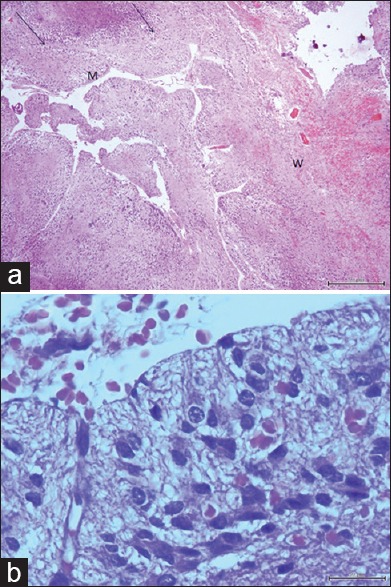
Figure 3.

(a) Microphotograph of tumor tissue reveals widened of cerebellar folia (black arrows) and thickened of the molecular layer (m) and thinned of central white matter (w) (H and E, × 40). (b) Microphotograph of tumor tissue shows numerous of dysplastic ganglion cells (H and E, × 400)
