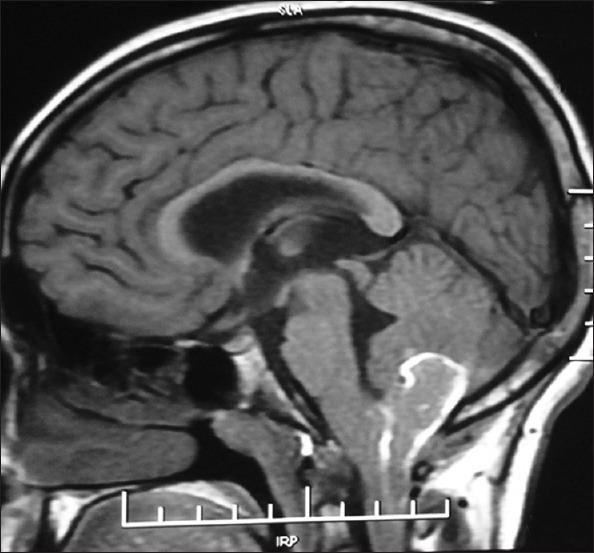
Figure 2.

Plain magnetic resonance imaging of brain, T1Wi showing mid posterior cerebellar, sub-acute, intracerebellar bleed with extension down towards foramen of Magnum. Peripheral hyperintensity in plain T1Wi image is due to peripheral meth haemoglobin component
