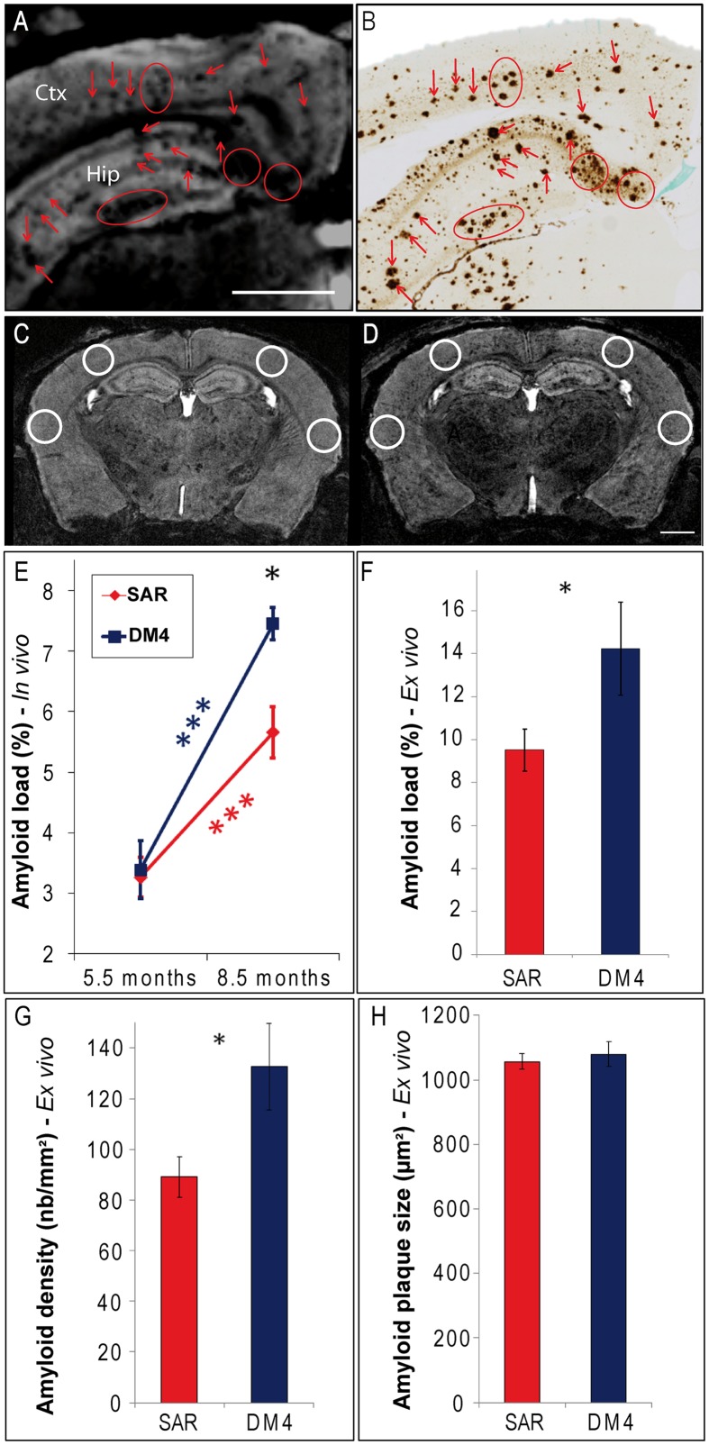
FIGURE 1.
Modulation of amyloid load following immunotherapy with SAR255952 or DM4 monoclonal antibodies. Registration between MR images (A) and amyloid-stained histological sections (B; 6E10 immunohistochemistry) in an 8.5-months-old APP/PS1 mouse. Hypointense spots observed on the MR images at the level of the cortex (Ctx) and hippocampus (Hip; A) are localized with amyloid plaques detected on the histological sections (B). ROIs used for plaque counting displayed on MR images of an APP/PS1 mouse at the age of 5.5 (C) and 8.5 months [D; A/P level = -2.2 mm compared to the Bregma (Paxinos and Franklin, 2001)]. (E) Measures from MR sections revealed an increased amyloid load between 5.5 and 8.5 months in SAR255952 and DM4-treated animals (repeated measure ANOVA and post hoc analysis within each group F[1,11] = 23 and 29, respectively, ∗∗∗p < 0.001). At 8.5 months, the amyloid load was lower in SAR255952-treated animals (n = 9) compared to control DM4-treated mice (n = 4; repeated measure ANOVA and post hoc analysis F[1,11] = 7, ∗p = 0.02). Histological measures confirmed the lower amyloid load [F, Student’s t-test, t(10) = 2.3, ∗p = 0.04] and reduced density of amyloid plaques [G, t(10) = 2.7, ∗p = 0.02] in SAR255952-treated animals at 8.5 months. The average size of the amyloid plaques was not modulated by therapy [H, t(10) = 0.6, ns]. Amyloid load in (E,F) is expressed as the proportion (%) of tissue area occupied by hypointense spots (E) or 6E10 immunoreactivity (F). Scale bars: 1 mm. Error bars represent standard error of the mean.

