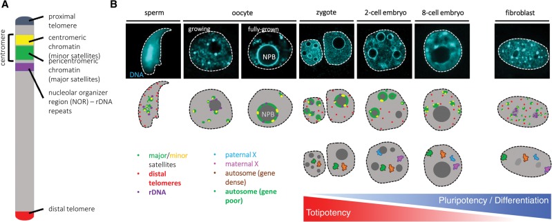
Figure 1.
Changes of chromatin organization during preimplantation development. (A) Schematic representation of the main building blocks and their organization on a mouse chromosome. rDNA repeats are present in only five of the 20 chromosomes in mice. (B) The differences in nuclear organization between mature gametes, preimplantation stages, and somatic cells are readily observable by a simple DNA (DAPI) staining. The top part of the scheme depicts the localization of the repetitive DNA elements studied by FISH: major satellites (green), minor satellites (yellow), rDNA (purple), and distal telomeres (red). In the mature sperm, rDNA and centromeric repeats cluster in the center, while distal telomeres are peripheral. During oocyte growth, centromeric repeats gradually assemble into a ring-like structure around the nucleolar precursor bodies (NPBs) and stay associated with them until the late two-cell stage, when they start reclustering into chromocenters, similar to somatic cells (fibroblast). rDNA is associated with the periphery of NPBs in oocytes and early embryos and gradually occupies nucleolar localization as somatic-like nucleoli form in place of NPBs. The localization of telomeres has not been determined in oocytes and, apart from the mature sperm, their localization has not been associated with nuclear compartments. The second scheme shows the localization of gene-rich (orange) and gene-poor (green) chromosomes as well as paternal (light blue) and maternal (pink) X chromosomes. Based on studies in bovine embryos, the gene density of a chromosome does not correlate with its radial position prior to embryonic genome activation (EGA, which occurs at the eight-cell stage in cattle and at the two-cell stage in mice). However, after EGA, gene-poor chromosomes occupy more peripheral localization compared with gene-dense ones. The imprinted inactive paternal X chromosome is preferably associated with the NPB in two- and four-cell stage mouse embryos but relocates from the NPB by the eight-cell stage. In the somatic cell depicted, due to random X inactivation, one of the X chromosomes forms the Barr body, preferentially located at the nuclear periphery.

