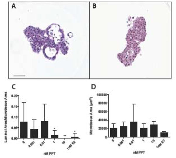
Figure 2.

Selective estrogen receptor alpha activation by PPT disrupts MCF-7 microtissue morphology. Representative histological sections of 3D MCF-7 microtissues grown in vehicle control (A) or 5nM PPT (B) for 7 days. PPT treatment reduced the luminal formation within MCF-7 microtissues (C), and did not alter microtissue area (D). Data is displayed as mean ± SEM, with analysis using a one-way ANOVA. Scale bar = 50μm. * p<0.05, **p<0.01
