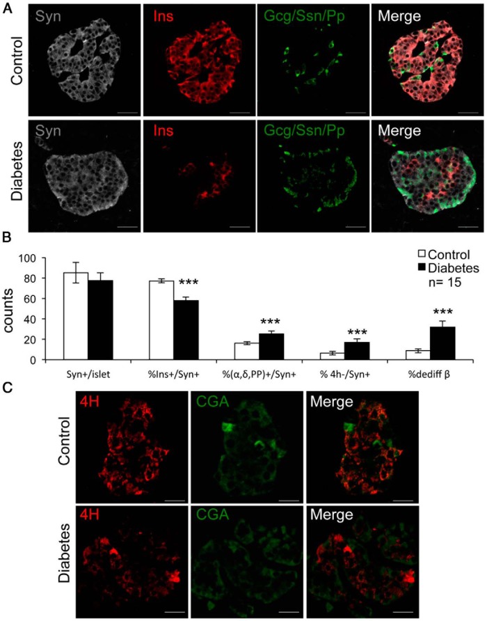
Figure 1.
Representative images of dedifferentiated β-cells. A, Immunofluorescent histochemistry on pancreatic section using insulin (Ins) (red), combined Gcg, Ssn, PP (green), and Syn (gray). B, Quantitative analysis of the data in A. C, Immunofluorescent histochemistry with the 4-hormone cocktail (4H) (red) and chromogranin A (CGA) (green). Data in B are mean ± SEM. ***, P < .001 by Student's t test. Scale bars, 20 μm; n = 15 for each group.

