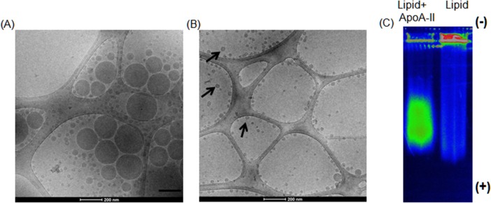
Fig 1. ApoA-II reduces the size of lipid.
Cryo-TEM micrograph picture of the SMOFlipid emulsion without ApoA-II (A) and after addition of ApoA-II (B). Note the bi-layer structure of the lipid surface of the nanoparticle like structures in presence of ApoA-II (black arrows). (C) Spectral flourescence color photograph of a 0.5% agarose gel run for 45 min at 90 V in 1 × TBE buffer. SMOFlipid labeled with DiD did not migrate through the well but reconstituted labeled SMOFlipid with ApoA-II migrated as a band.

