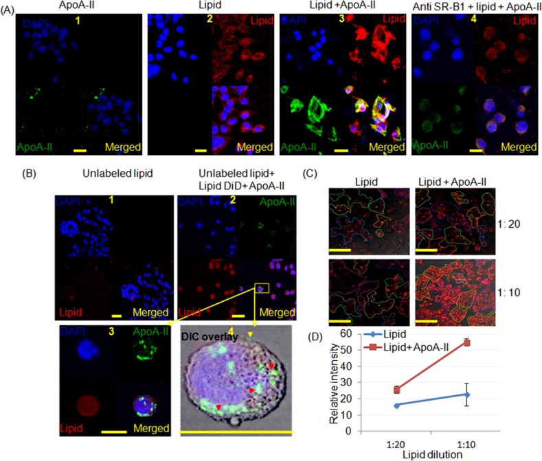
Fig 2. Confocal micrographs of CFPAC-1 cells demonstrating the influence of ApoA-II on lipid uptake.
SMOFlipid was labelled with DiD (red), ApoA-II was labelled green with a secondary antibody and DAPI stained the nuclei blue. (A1) cells treated with ApoA-II alone, (A2) cells treated with lipid alone, (A3) cells treated with reconstituted lipoproteins after adding ApoA-II to DiD labeled SMOFlipid (SMOF/A-II) demonstrating increased uptake and (A4) cells treated with (SMOF/A-II) after pretreating with anti-SR-B1antibodies which reduced the lipid uptake (scale bar, 20 μm). (B) Cells were pretreated with unlabeled lipid at 1:10 dilution and (B2) after 2 h SMOF/A-II was added, it appeared to be endocytosed in cytoplasmic vesicles as shown in (B3) and (B4). ApoA-II (green) is attached with the cell membrane (yellow arrow head) and as well in the cytoplasm in part associated with the lipid (red arrow head) in Differential Interference Contrast (DIC) overlay image (scale bar 25 μm). (C) Live cell imaging experiment demonstrated greater uptake of lipid in cells when reconstituted lipoproteins after adding ApoA-II to DiD labeled SMOFlipid was added to the media for lipid concentrations of 1:20 and 1:10 (scale bar 100 μm). (D) The intensity of the DiD staining was greater when 4 samples were examined with eight to twelve areas of interest for each study where ApoA2 increased the uptake of lipid 25.6±2.2, P = 0.02 and 54.8±2.2, P = 0.004 for dilutions of 1:20 and 1:10 respectively.

