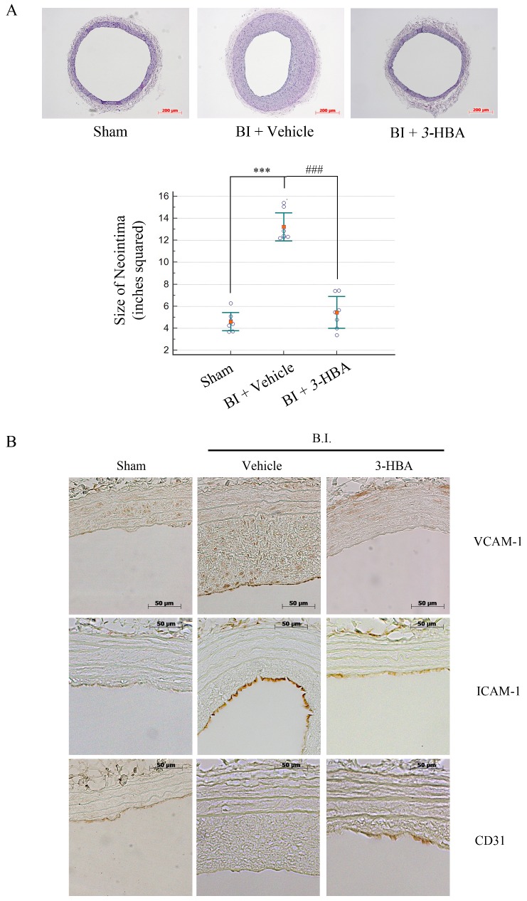
Fig 6. The effect of 3-HBA in CCA balloon-injured Sprague Dawley rats.
(A, B) Seven-week-old SD rats (200 g) were treated with the appropriate substance by i.p. injection for 2 weeks, and then common carotid arteries (CCAs) from the rats were balloon-injured. The chosen substance was injected for another 4 weeks, and then the rats were sacrificed for (A) hematoxylin and eosin staining of CCAs. The graph shows the percentage of neointima area in SD rats from each group (n = 7). Measurements were performed using Scion Image software. (B) Immunohistochemistry staining of rat aortas show CD31, VCAM-1, and ICAM-1 production in the linings of the aorta from the same tissues used in Fig 6A. Data are presented as the mean ± SEM. Values represent the mean ± SEM of three experiments; *** indicates p < 0.001 compared to the sham group. ### indicates p < 0.001 compared to the vehicle group.

