FIGURE 1:
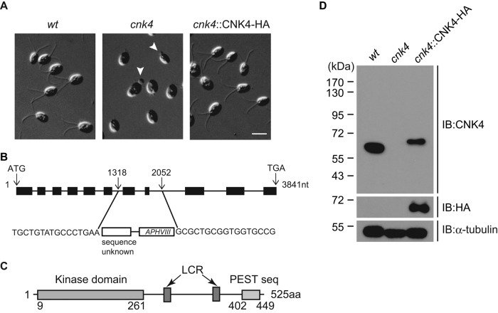
cnk4 is defective in ciliogenesis. (A) Wild-type (wt), cnk4, and cnk4::CNK4-HA cells were imaged by DIC microscopy. Note that cnk4 cells are aflagellated or have flagellar buds (arrowheads). Bar, 10 μm. (B) Schematic illustration of the CNK4 gene, showing the replacement of exons 7 and 8 by foreign DNA fragment during random DNA insertion. The insertion site was identified by PCR and DNA sequencing. (C) The domain structure of CNK4. Numbers are amino acid positions. LCR, low-complexity region. (D) Western blot of whole-cell lysates from indicated strains with antibodies against CNK4, HA, and α-tubulin. No CNK4 was detected in the cnk4 mutant.

