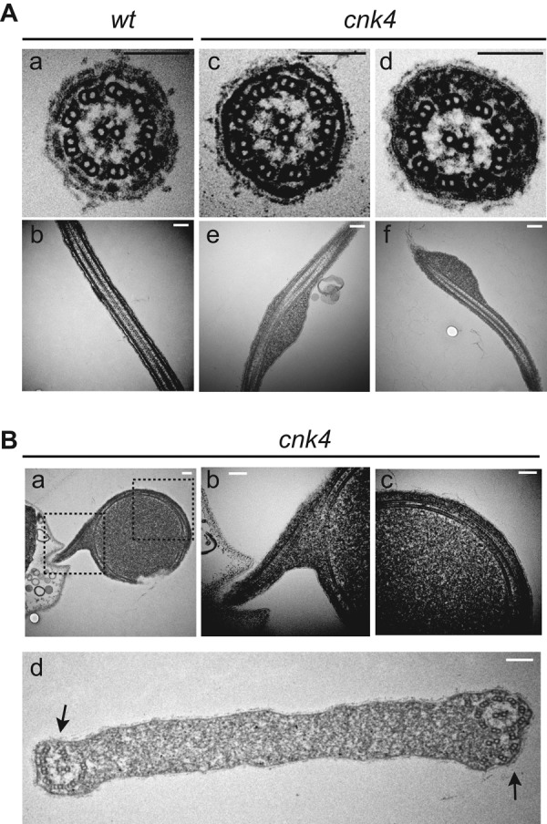
FIGURE 5:

Electron microscopic analysis of cnk4 flagella. (A) Cross- and longitudinal sections of wt (a, b) and cnk4 (c–f) flagella. Note that cnk4 flagella accumulate amorphous materials between the membrane and the axoneme (d–f), whereas its substructures appear normal (d). Bars, 150 nm. (B) Flagellar balloon cross-sectioned to show curled axoneme (a–d) and degenerated axonemal microtubules (d, arrows). Dashed box images in (a) are enlarged and shown in (b) and (c), respectively. Bars, 150 nm.
