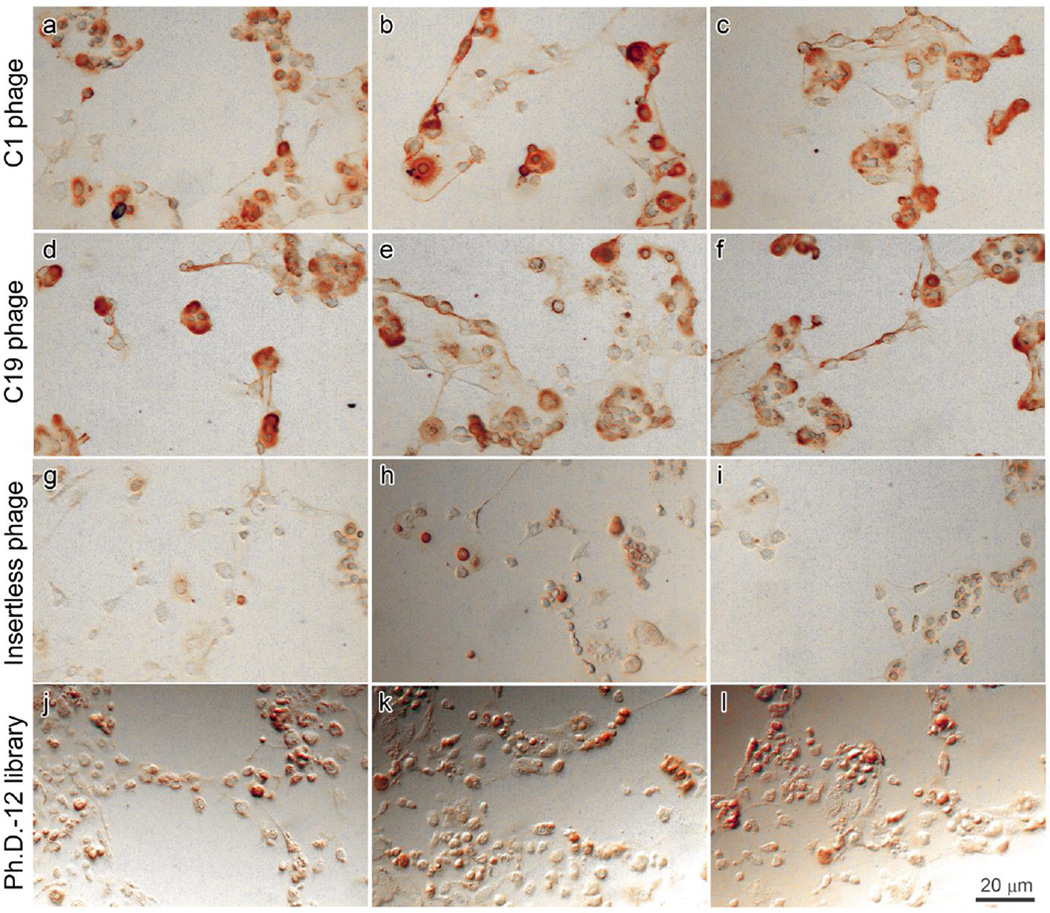
Figure 3.
Localization of phage binding to cultured chondrocytes by immunohistochemistry. C1 phage (a–c), C19 phage (d–f), insertless phage (g–i), and naive phage library (j–l) were incubated with primary culture of mouse epiphyseal chondrocytes, detected by HRP-conjugated anti-M13 antibody and stained using DAB substrate to produce a brown color. Images were taken from three representative fields.

