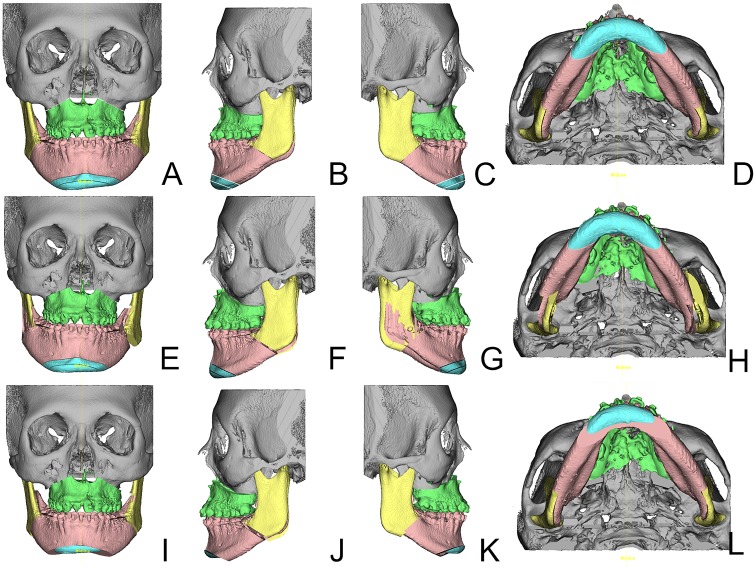
Fig 3. Visualization of the three positions in sample patient #25 (Table 1).
Isolated cleft palate patient. From left to right frontal, left lateral, right lateral and basal views are shown of the same position. Subfigures A-D show the initial Position0, Subfigures E-H the result of the 2D plan translation into the 3D environment and Subfigures I-L the adjusted Position2 in the 3D plan agreed upon by the orthodontists and the surgical team. Note the severe bony collision in the right ramus area of this patient in Subfigures E and G and the subsequent large bony gap in the left ramus area. Through counterclockwise yaw rotation the collision and overall asymmetry were resolved (Subfigures H and L, E and I). The genioplasty position was altered by shortening the chin segment, thus reducing the patient’s facial height (Subfigures I-L).

