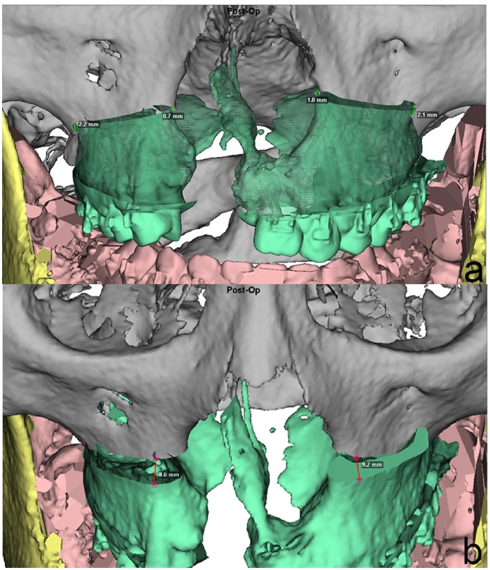
Fig 8. Position analysis of the Le Fort I segment of sample patient #29 (Table 1).
Bilateral cleft lip/palate patient. The 3D position of the Le Fort I segment in relation to the superior fixed part of the maxilla is recorded in both lateral and piriform areas to facilitate proper positioning of the MMC during surgery. Midline shifts in the patient’s right lateral pillar and impaction of the left piriform aperture (Fig 5a) are noted as well as the different amounts of advancement or setback in both left and right lateral and piriform levels (Fig 5b). This information is the result of the 3D alternation of the MMC and helps to transfer the results of 3D simulation into the surgical setting.

