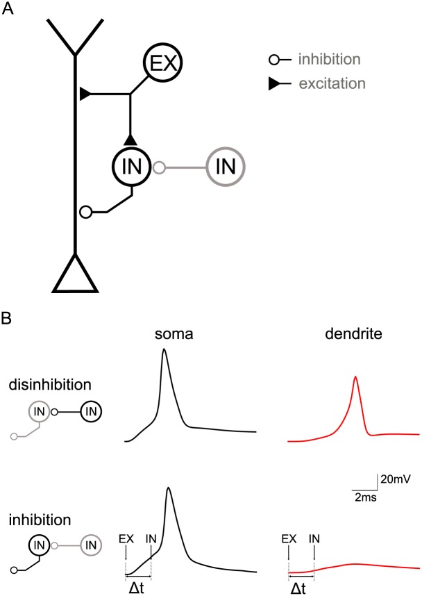
Fig 7. Circuit model of feedforward inhibition.
A: Topology of the circuit with a multi-compartment pyramidal neuron model and a model of a fast-spiking inhibitory interneuron (IN, targeting pyramidal neuron) receiving excitatory input from a source (EX) at time t0 and potentially tonic disinhibition (IN, targeting IN). B: Effect of switching the feedforward interneuron on and off (via a second interneuron—marked gray in panel A) measured in the dendrite of the pyramidal neuron (370 μm from the soma), which in turn is driven by apical excitation. Note that the circuit is identical to that in panel A, although the lefthand schematics only zoom in on the interneurons. Upper paradigm: When the feedforward interneuron is switched off (in gray) because of activation of the second interneuron (in black), excitation triggers a spike in the pyramidal cell’s soma which propagates unhindered into the dendrite. Lower paradigm: When the feedforward interneuron is active (in black) due to excitation and because the second interneuron is switched off (in gray), excitation triggers a spike in the pyramidal cell’s soma that does not propagate far into the dendrite due to the inhibition provided by the feedforward interneuron (90 μm from soma, Δt = 2 ms).

