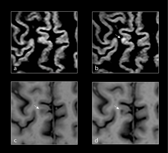
Fig 2. Reclassification of new cortical lesions.
Baseline and follow up axial DIR (a, b) of the brain of a patient with primary progressive multiple sclerosis (PPMS) demonstrating a new focal lesion in the cortical grey matter (white-arrows) at follow-up. Corresponding baseline and follow-up axial PSIR (c, d) of the same patient with PPMS demonstrating focal lesions in the cortical grey matter (white-arrows) at both baseline and follow-up.

