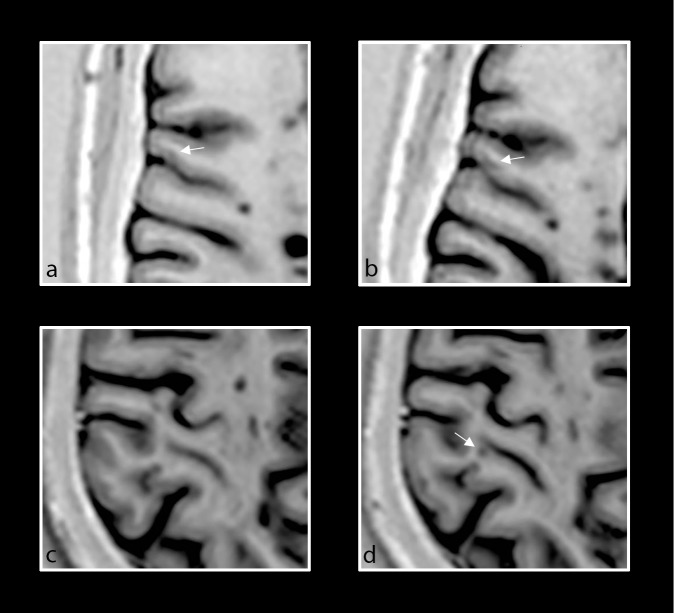
Fig 3. Retrospective analysis of new PSIR cortical lesions.
Baseline and follow-up axial PSIR images of different patients with primary progressive MS demonstrating focal lesions in the cortical grey matter. Intracortical lesion that was too small to be counted at baseline (a, white arrow), but that enlarged and was counted on follow-up scan (b, white arrow). New LC lesion (d, white arrow) noted at follow-up but not at baseline (c).

