
Figure 3.
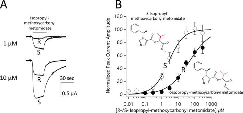
Direct activation of α1(L264T)β3γ2 γ-aminobutyric acid type A (GABAA) receptors expressed in oocytes by R-isopropyl-methoxycarbonyl metomidate and S-isopropyl-methoxycarbonyl metomidate. (A) Electrophysiological traces recorded upon perfusing an oocyte expressing α1(L264T)β3γ2 GABAA receptors with 1 μM (top) or 10 μM (bottom) S- or R-isopropyl-methoxycarbonyl metomidate. The same oocyte was used to obtain all 4 traces. (B) S- and R-isopropyl-methoxycarbonyl metomidate concentration-response relationships for α1(L264T)β3γ2 GABAA receptor direct activation. Each data point is the mean value (± SD) obtained from 6 different oocytes. In each panel, the chemical structures are shown as insets with the differences between analogues highlighted in red. The curves are fits of the two data sets to a Hill equation yielding half-maximal direct activating concentrations of 2.6 ± 3 μM and 46 ± 6 μM for S- and R-isopropyl-methoxycarbonyl metomidate, respectively (p<0.0001).