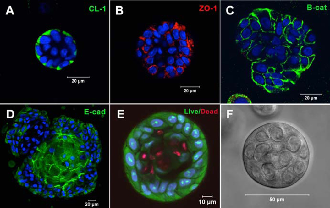
Figure 5.
Acini-like spheroids in 3D HA hydrogels. Spheroid structures express tight junction markers CL-1 (A), ZO-1 (B), E-cadherin (D) and adherens junction marker, β-catenin (C). (E): Live/Dead staining of Syto13 positive green cells and propidium iodide positive red cells. Nuclei stain blue. (F): Representative phase image of an acinus-like structure. Reprinted from Pradhan-Bhatt et al, 201315 with permission.

