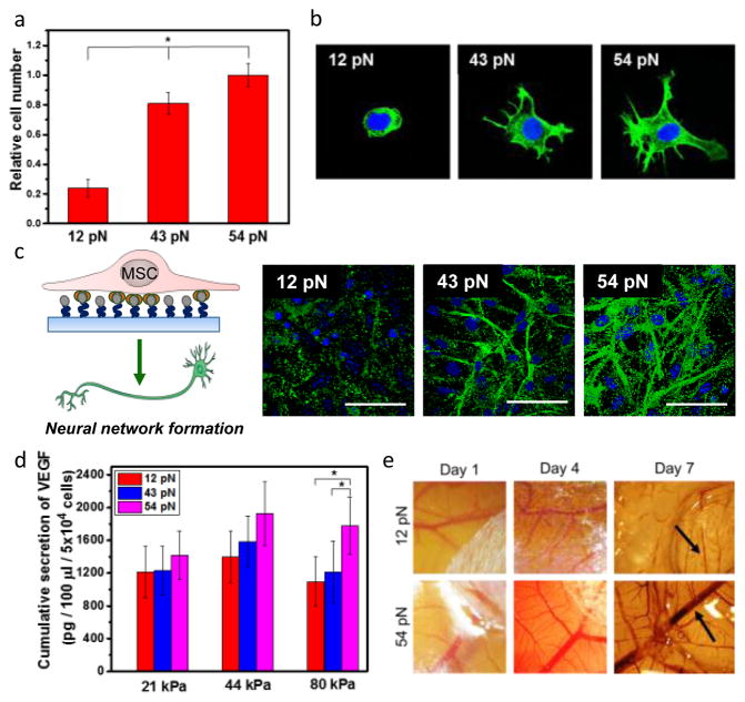
Figure 3. Analysis of the role of Ttol of RGD-DNA tether on adhesion and phenotypic activities of MSCs.
a, Quantification of the number of MSCs adhered to a hydrogel modified with RGD-DNA tether with controlled Ttol. b, Fluorescence image of F-actin (green) of MSCs. c, Analysis of neural differentiation of MSCs. Differentiated neural cells were positively stained for MAP-2 (green). d, The total amount of VEGF secreted by MSCs cultured on alginate-g-biotin hydrogels with different elastic moduli modified with RGD-DNA tether with controlled Ttol. e, Microvascular networks of the CAM implanted with the MSC-hydrogel constructs. Arrows indicate blood vessels. The error bars represent the standard deviation of three measurements. * indicates statistical significance of the difference (*p < 0.05).

