Abstract
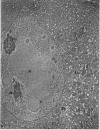
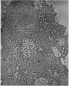
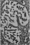
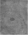
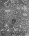
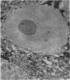
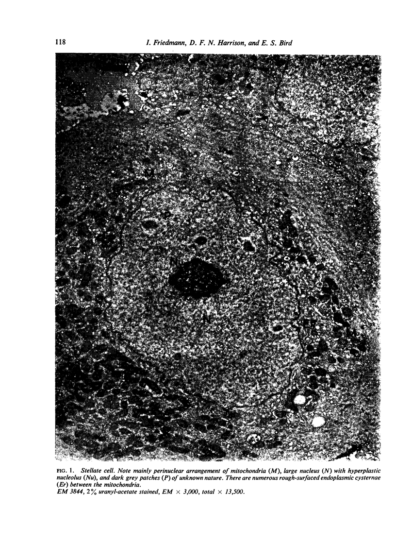
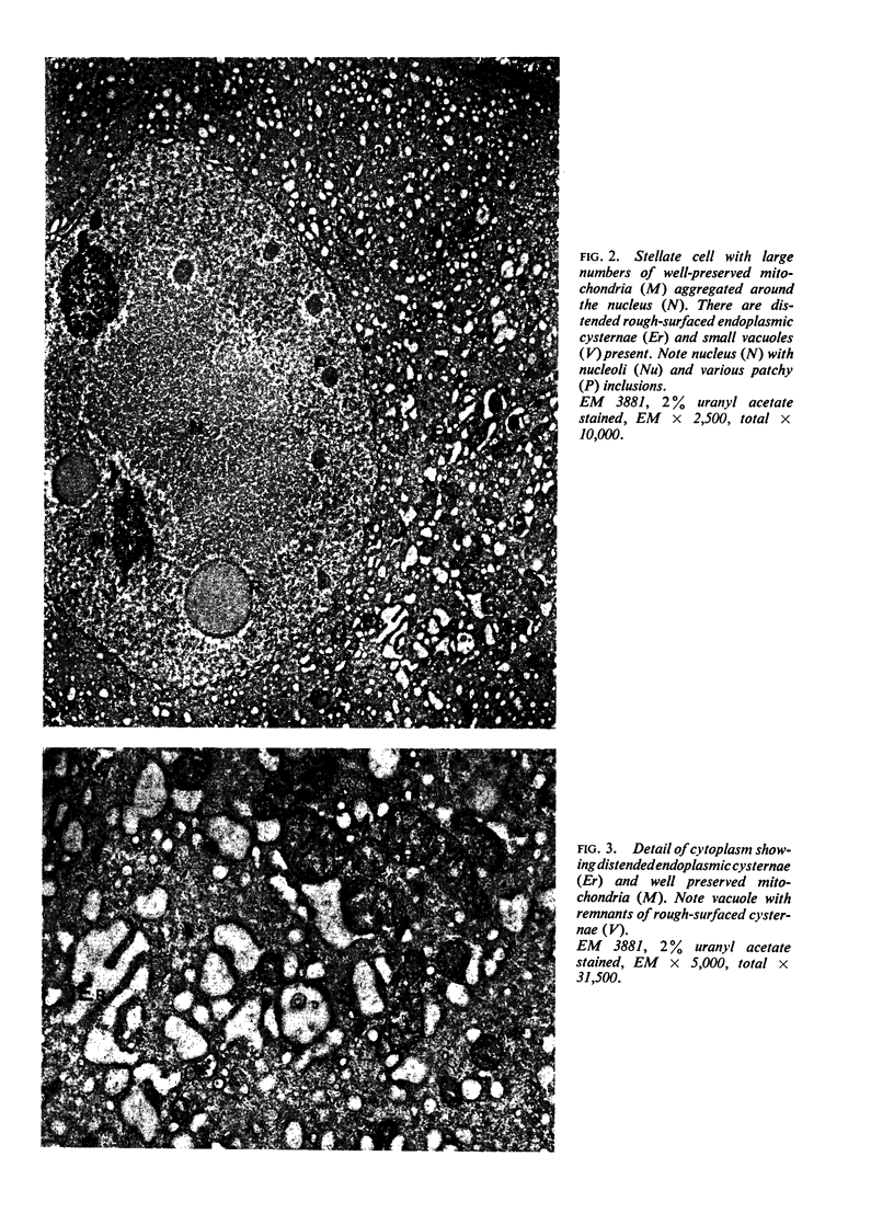
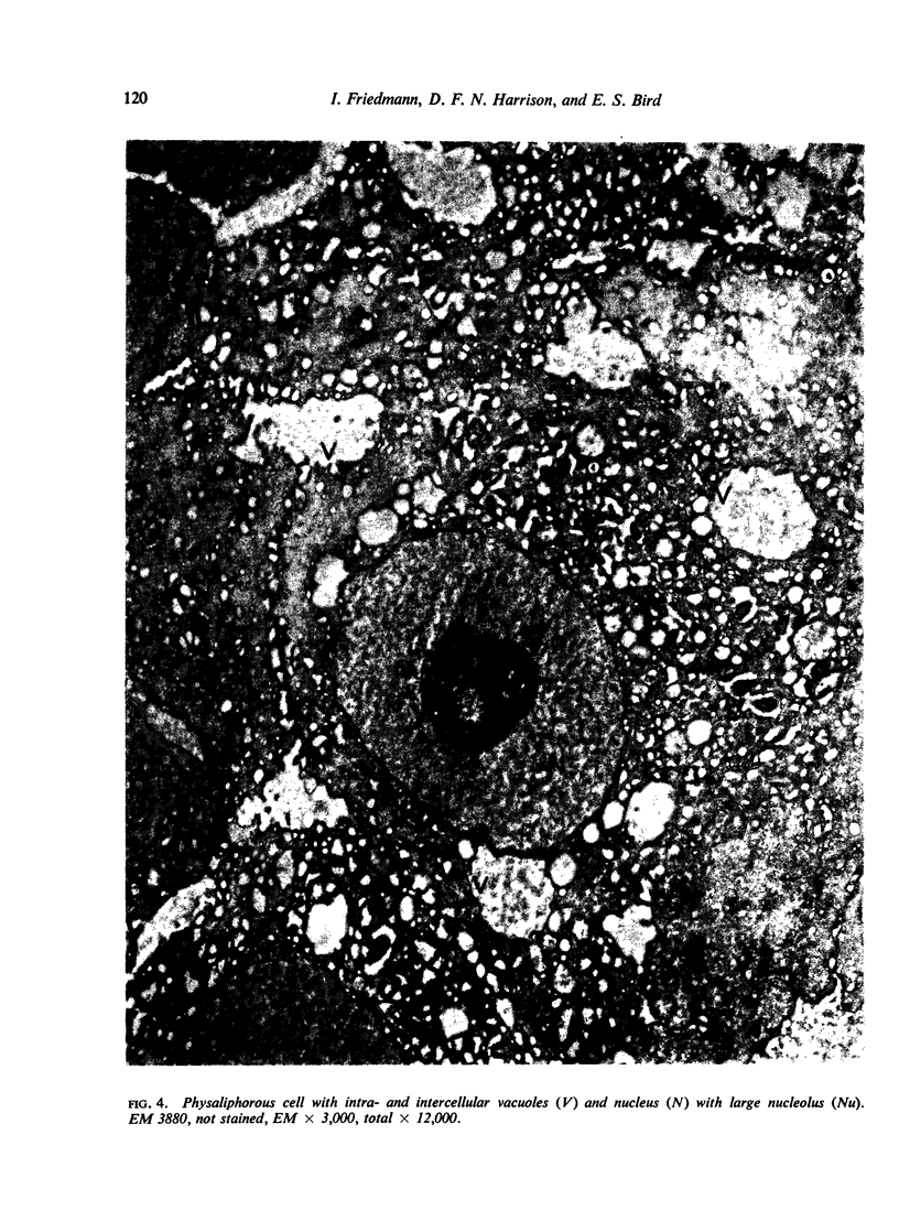
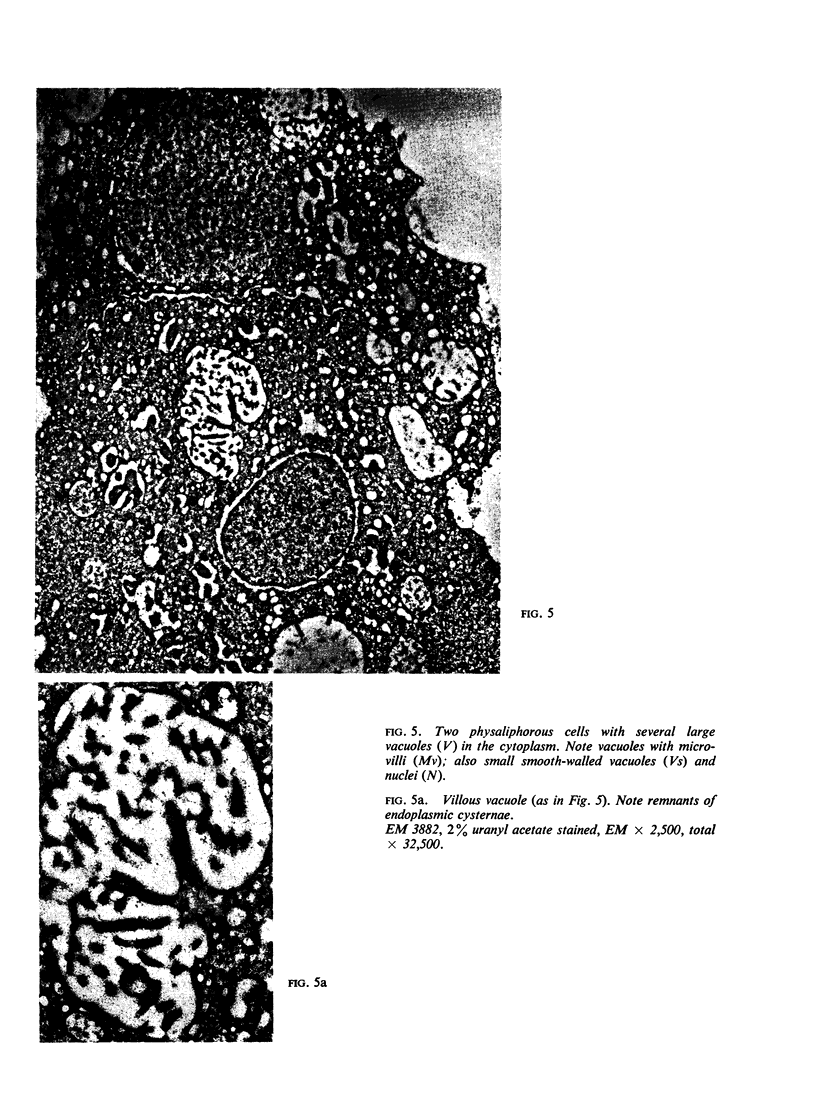
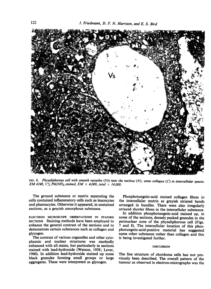
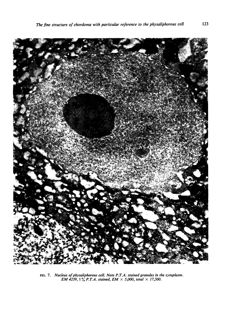
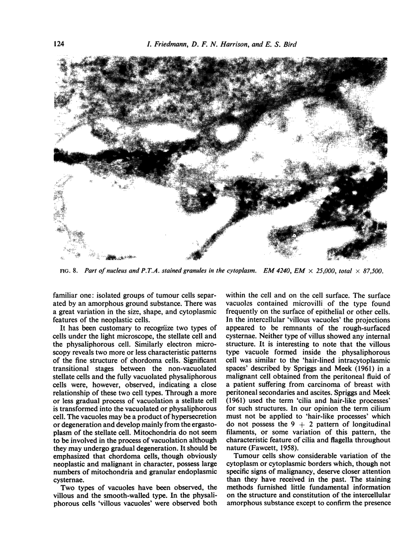
A chordoma was examined by electron microscopy. It was composed of stellate and physaliphorous cells. The fine structure of these cells is described. Transitional type cells are also described and it is suggested that the physaliphorous cells develop from the stellate cells through a process of vacuolation. Two types of vacuoles are described, `smooth-walled' and `villous' endowed with numerous structureless microvilli. Some observations on stained sections are discussed.
Full text
PDF
Images in this article
Selected References
These references are in PubMed. This may not be the complete list of references from this article.
- DROCHMANS P. [Demonstration of glycogen in liver cells by electron microscopy]. J Biophys Biochem Cytol. 1960 Oct;8:553–558. [PMC free article] [PubMed] [Google Scholar]
- HARRISON D. F. A case of primary chordoma of the sphenoidal sinus. J Laryngol Otol. 1961 Apr;75:429–432. doi: 10.1017/s0022215100057911. [DOI] [PubMed] [Google Scholar]
- IURATO S., LEONARDELLI G. B. Il profilo istomorfologico e istochimico del cordoma rinofaringeo. Tumori. 1956 Jul-Aug;42(4):559–575. doi: 10.1177/030089165604200404. [DOI] [PubMed] [Google Scholar]
- LEVER J. D. A method of staining sectioned tissues with lead for electron microscopy. Nature. 1960 Jun 4;186:810–811. doi: 10.1038/186810a0. [DOI] [PubMed] [Google Scholar]
- ORMEROD R. A case of chordoma presenting in the nasopharynx. J Laryngol Otol. 1960 Apr;74:245–254. doi: 10.1017/s0022215100056528. [DOI] [PubMed] [Google Scholar]
- WATSON M. L. Staining of tissue sections for electron microscopy with heavy metals. II. Application of solutions containing lead and barium. J Biophys Biochem Cytol. 1958 Nov 25;4(6):727–730. doi: 10.1083/jcb.4.6.727. [DOI] [PMC free article] [PubMed] [Google Scholar]


