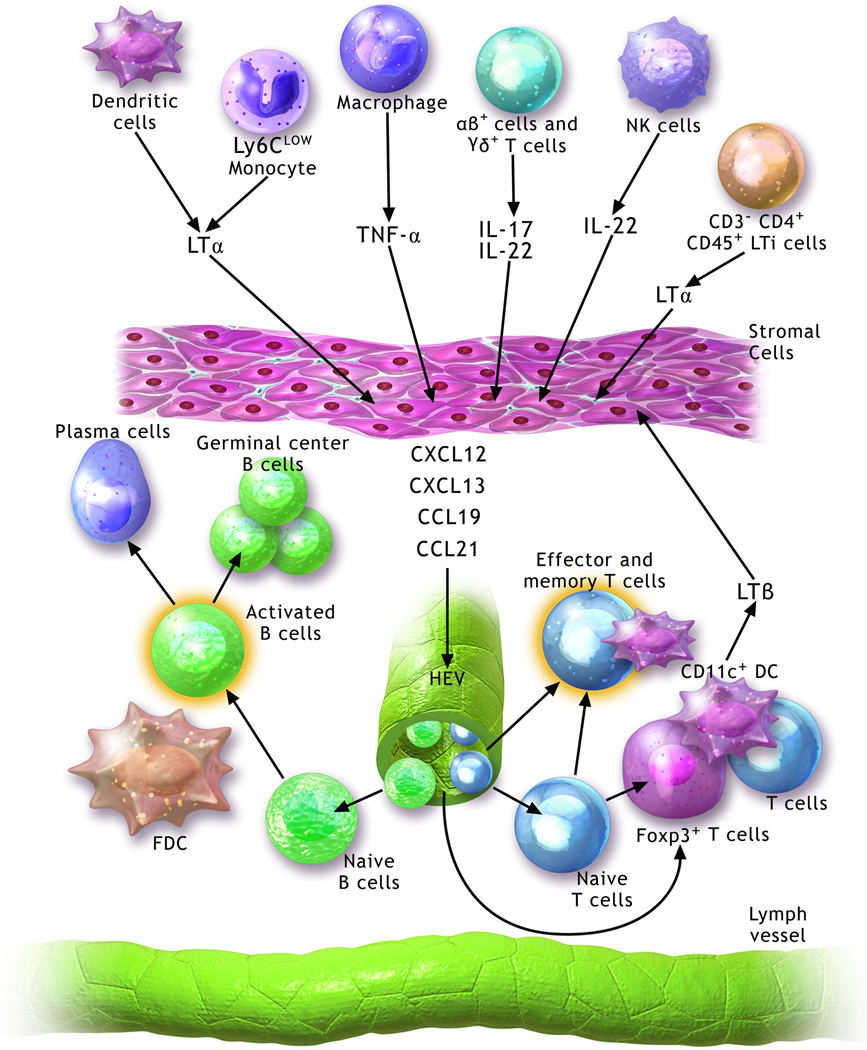
Figure 1. Molecular and cellular pathways contributing to the development and maintenance of tertiary lymphoid organs.
Several cell populations including lymphoid organ inducer cells (LTi) (CD3−CD4+CD45+), natural killer (NK) cells, dendritic cells (DC), macrophages and Ly6Clow monocytes may contribute to the development of TLOs through the production of various mediators, including lymphotoxin (LT)-α, tumor necrosis factor (TNF)-α, IL-17 and IL-22. These activate stromal cells to secrete homeostatic chemokines (CCL19, CCL21, CXCL12 and CXCL13), which promote the recruitment of B cells and T cells. A well-organized tertiary lymphoid organ is composed of separated T and B cell areas, follicular dendritic cells (FDC), CD11c+ dendritic cells, high endothelial venules (HEVs) and lymphatic channels. CD11c+ dendritic cells contribute to the maintenance of these structures possibly through secretion of LTβ.

