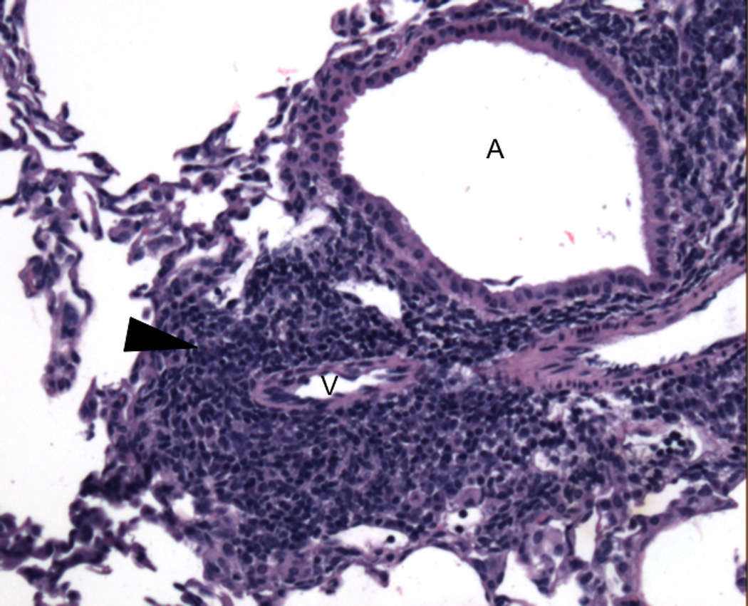
Figure 2. Tertiary lymphoid organ in accepted mouse lung allograft.

Representative H&E photomicrograph of bronchus associated lymphoid tissue (arrowhead) in an accepted BALB/c lung allograft 30 days after transplantation into an immunosuppressed B6 recipient (MR1 250 µg intraperitoneally day 0, CTLA4-Ig 200 µg intraperitoneally day 2). A: airway; V: vessel. Picture was taken on an Olympus BX51 imaging system under the 10× objective lens (100× total magnification) with an AmScope MT1400-CCD camera.
