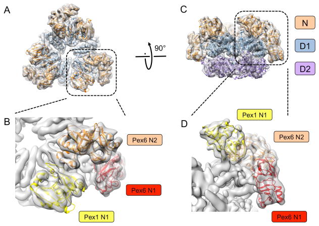
Fig. 1. Cryo-EM structure of the Pex1/Pex6 complex.
(A) Models of Pex1 and Pex6 were fitted into the density map (grey envelope) determined in the presence of ATPγS without symmetry imposed [22]. Shown is a top view with the D1 domains colored in blue and the N domains in brown. (B) Magnified view of the N domains in (A). The N domains are distinguished by different colors. (C) As in (A), but side view and including the D2 ring (purple). (D) Magnified view of the N domains in (C), colored as in (B).

