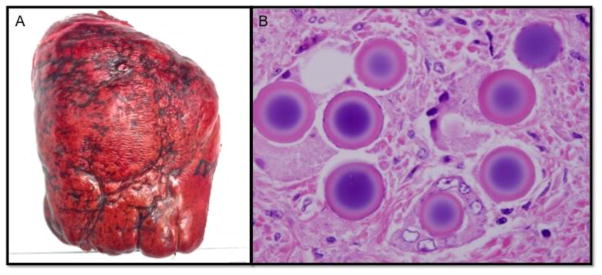
Figure.
(A) Resected liver segments IV–VIII demonstrate significant Y90-induced radiation fibrosis and damage that is unique to post radioembolized liver and complicates resection. (B) Histopathologic sections revealed amorphous microspheres ranging from 25 to 55 microns in diameter generally within vascular spaces and usually in clusters. Oil immersion revealed the microspheres to demonstrate a refractile appearance and to have a basophilic center that often was surrounded by a white halo with an eosinophilic periphery.

