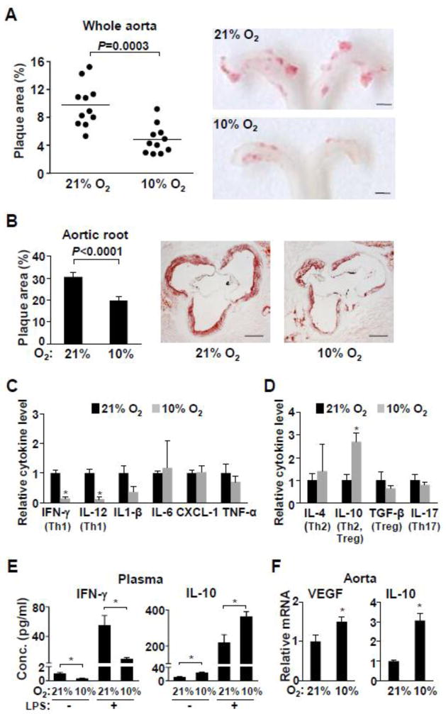
Fig. 1.
Low ambient oxygen reduces atherosclerosis and inflammation in ApoE−/− mice. ApoE−/− mice (5–6 wk old) were maintained in room air (21% O2) or placed in a hypoxia chamber (10% O2) for 22 wk prior to analyses (except for 1e). a The aortas were longitudinally dissected and atherosclerotic plaque areas (red) were quantified after staining with Sudan IV. Shown are representative images of the aortic arch where most of the plaques develop (n=11). Scale bars: 1 mm. b Aortic root cross sections were stained with Oil Red O and the stained plaque areas were measured (n=10–11). Representative aortic root sections are shown. Scale bars: 200 μm. c ApoE−/− mice were exposed to the indicated ambient oxygen for 22 wk and the proinflammatory cytokine level in plasma were measured by multiplex immunoassay kit (Meso Scale Discovery) (n=5). Cytokines that can serve as Th1 markers are indicated in brackets. d Levels of plasma cytokines that can serve as markers of other Th subtypes (indicated in brackets) were measured by individual immunoassay kits (Thermo and eBioscience) (n=5). e Effect of hypoxia on both basal and LPS-stimulated plasma cytokine levels in ApoE−/− mice. These mice were exposed to the indicated ambient oxygen for 5 wk prior to i.p. injection of LPS (n=5–11). f Hypoxia responsive VEGF and anti-inflammatory cytokine IL10 gene expression in aortic tissue were measured by real-time RT-PCR (n=6). Values shown as mean ± SEM, *p < 0.05.

