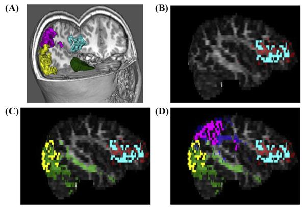
Fig. 2.
Example of merging multi-model imaging data for probabilistic tractography. (A) Cortical gray matter used in tractography are displayed in FSL’s 3D viewer in a color-coded fashion (green = RAC mask; yellow = right OT mask; pink = right TP mask; blue = right IFG mask). (B) Cortical GM mask of left IFG from Freesurfer is co-registered with DTI ProbtrackX output for evaluating structural connectivity between RAC and Left IFG (red). (C) Cortical GM mask of left OT mask from Freesurfer is co-registered with DTI ProbtrackX output for evaluating structural connectivity between RAC and left OT (green). (D) Cortical GM mask of left TP mask from Freesurfer is co-registered with DTI ProbtrackX output for evaluating structural connectivity between RAC and left TP (blue).

