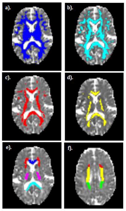
Figure 1.
Examples of ROIs used in this study displayed on a single subject. a–d). FA thresholded ROIs. e–f). Anatomical WM ROIs. a). FA > 0.3 b). FA > 0.4 c). FA > 0.5 d). FA > 0.6. e). Various anatomical WM ROIs including genu (blue), splenium (cyan), posterior limb of the internal capsule (purple), and the anterior corona radiata (red). f). Same as e), but a more superior slice to show the remaining ROIs including the superior corona radiata (yellow) and the posterior corona radiata (green). The red ROI is a continuation of the anterior corona radiata as seen in e).

