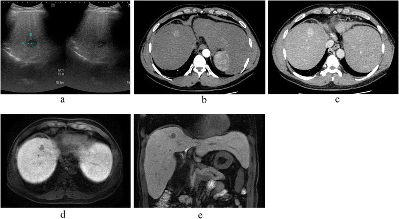
Fig. 1.

Preoperative radiological findings. a Abdominal ultrasonography showed a hypoechoic lesion in segment 8 (S8) of the liver. b The main tumor (S8) showed an early enhancement in CT, which was prolonged until the delayed phase (c). d Gadolinium ethoxybenzyl diethylenetriaminepentaacetic acid (Gd-EOB-EDTA)-enhanced MRI revealed a solitary 1.8-cm tumor, with a low-intensity signal in the hepatobiliary phase in the transverse (d) and sagittal planes (e)
