
Figure 8.
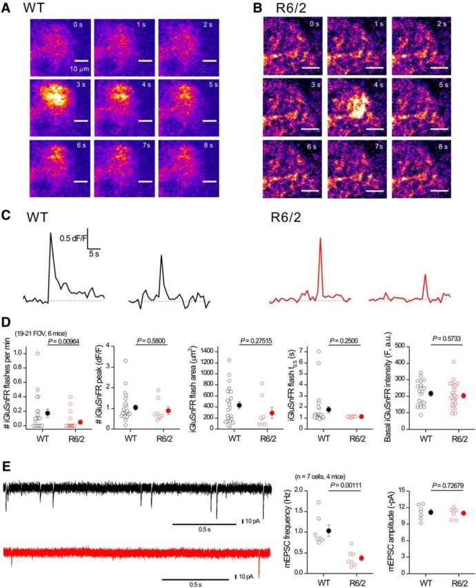
iGluSnFR flashes are not increased in striatal astrocytes from R6/2 mice. A, Representative time series of confocal images from a small region of an astrocyte expressing iGluSnFR from a WT mouse; an iGluSnFR flash occurred at ∼3 s. B, As in A, but for an astrocyte from a R6/2 mouse. C, Representative traces for transients such as those shown in A and B. D, Bar graphs showing average data for traces such as those shown in C. Note: the only significant difference in iGluSnFR signals between WT and R6/2 mice was in the frequency; no other parameter was changed in R6/2 compared with WT astrocytes. E, Representative traces and average data for mEPSCs recorded from MSNs at −70 mV from WT and R6/2 mice. In some cases, the error bars signifying SEM are smaller than the symbols used.