
Figure 4.
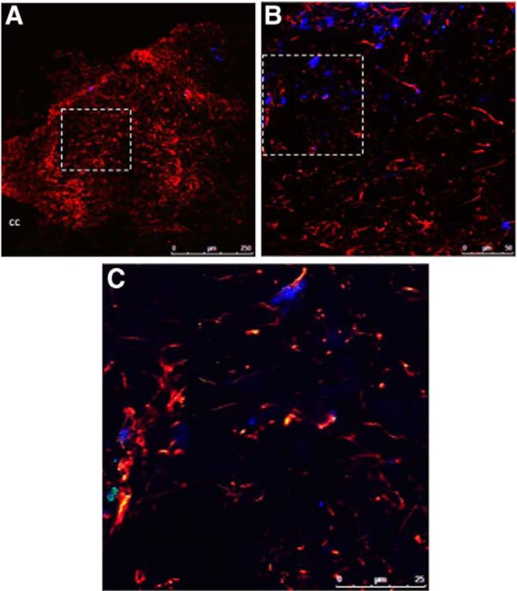
Fluorescently labeled Ex-4 is taken up by neurons and astrocytes within the NTS following fourth cerebroventricle administration. Rats (n = 3) were injected with fluoro-Ex-4 (0.3 μg, i.c.v.) and killed 3 h later. A, B, A representative NTS-containing brain section is shown at 20× magnification in A, with GFAP-positive cells shown in red and NeuN-positive cells in blue. The dotted box outlined in A is magnified in B. This higher-magnification image in B shows a single image within a z-stack (1 μm step size) taken with the 63× oil-immersion objective and depicts fluoro-Ex-4 labeling in GFAP-positive cells (red) and NeuN-positive cells (blue). The dotted rectangle indicates the location of an additional 2–3× optical zoom represented in C within the same z-stack (0.5 μm step size). cc, Central canal.