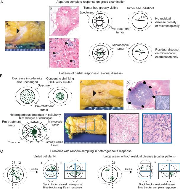
Figure 1.
Patterns of response in the breast and problems related to sampling for histologic evaluation: schematic overview with gross and microscopic illustrations. Photos courtesy of Veerle Bossuyt. (A) In some cases with complete response, a residual tumor bed is visible. In others, the tumor bed is indistinct and sampling of the correct area can only be confirmed by thorough clinical and imaging correlation and identification of a clip. Often, residual microscopic disease is identified when there is no residual tumor grossly. (a) Gross photograph of tumor bed (arrow). (b) Low-power hematoxylin and eosin (H&E) slide of this tumor bed. No residual tumor is identified. (c) High-power of H&E slide of the tumor bed from a different patient with rare residual invasive carcinoma cells (small arrows). (B) A partial response ranges from a decrease in cellularity with unchanged tumor size to concentric tumor shrinking with unchanged tumor cellularity. Often the decrease in cellularity is heterogeneous and residual disease extends beyond the grossly visible tumor bed. (a) Gross photograph of tumor bed with residual tumor (arrow). (b) H&E slides (low and high power) of different patient with residual invasive carcinoma concentrated in a nodule with high cellularity within the tumor bed (concentric shrinking). (c) Gross photograph of most common pattern of residual disease with scattered residual tumor across a fibrous tumor bed. (d,e) Medium power of H&E slides from two different blocks of the tumor bed (black and blue boxes) illustrating that cellularity often varies greatly from block to block. (C) When decrease in cellularity is heterogeneous, random sampling can lead to very different estimates of cellularity. When the decrease in cellularity is so heterogeneous that there are apparent areas with complete response (no residual disease) and apparent multiple foci of residual tumor (scatter pattern), there are interobserver variability and inconsistencies among guidelines in size measurements and when to consider multiple foci. For example, for AJCC staging, the largest contiguous focus of invasive carcinoma should be measured [23]. Intervening areas of fibrosis are specifically excluded, whereas other systems include these areas [24–26, 32, 33]. Moreover, there can be interobserver variability in how much fibrosis to allow within this largest contiguous focus.

