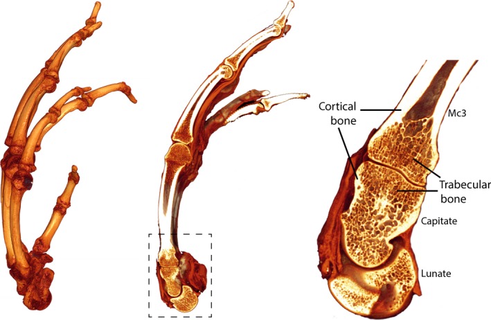
Figure 1.

Trabecular and cortical bone structure in a chimpanzee hand. A 3D rendering from a micro‐CT scan of an extant chimpanzee hand (Pan troglodytes; left), a sagittal cross‐section through the third ray, revealing the internal bone structure (middle) with the area outlined in the dashed box blown up (right) to show the dense cortical shell and the trabecular meshy network inside. Note that trabeculae fill just the epiphyses of long bones, like the third metacarpal (Mc3), while short bones, like the capitate and lunate, are filled completely with trabeculae.
