Figure 2.

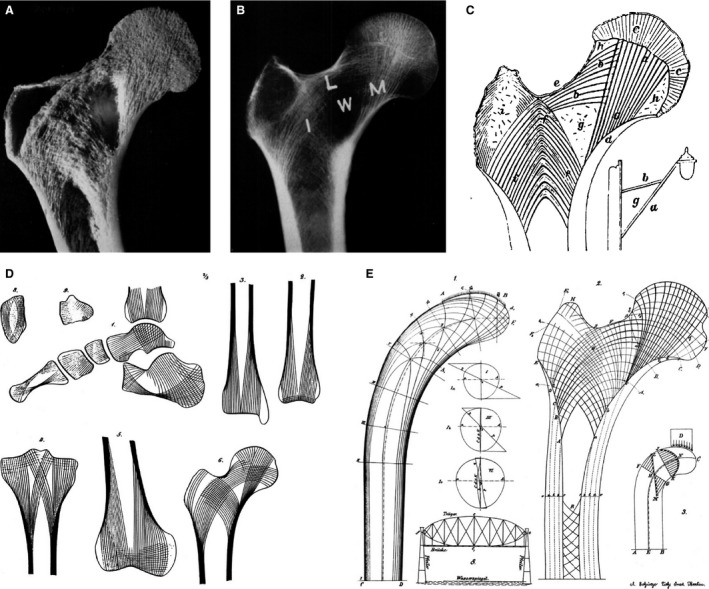
Historical description of trabecular bone functional adaptation. (A) Coronal cross‐section and (B) radiograph of a human proximal femur showing the distinct trabecular pattern related to bipedal loading. (C) Ward's (1838) drawing of the trabecular structure that he related structurally to the bracket street lamp post. The sparse area of trabeculae in the femoral neck is equivalent to the empty space within the bracket (‘g’), which is known as ‘Ward's triangle’ (W). (D) von Meyer's (1867) stylized illustrations of trabecular patterns in human bones. (E) Wolff's (1870) composite diagram including the compressive and tensile strain patterns in Culmann's cantilevered beam and ‘crane’ (left), and the similarity to the trabecular patter in the human proximal femur (right). Images (A–C) adapted from Garden (1961), and images (D and E) adapted from Skedros & Baucom (2007).
