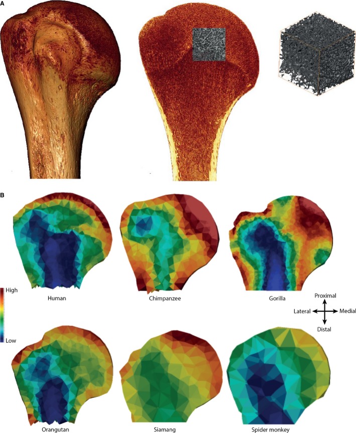
Figure 4.

Different methodological approaches to investigating trabecular bone. (A) The traditional volume of interest (VOI) method that analyses trabecular structure in a subsection of the epiphysis or bone. Here, an example is shown in the human proximal humerus in which the VOI is 30% of the geometric mean of the articular dimensions. (B) A new, holistic method [medtool (Pahr & Zysset, 2009a; Gross et al. 2014)] that quantifies and visualizes variation in trabecular bone distribution (BV/TV) and stiffness throughout the entire epiphysis or bone. Here, variation in BV/TV is shown in a coronal cross‐section throughout the proximal humerus in the same taxa and specimens shown in Fig. 3. Red indicates high BV/TV; blue indicates low BV/TV.
