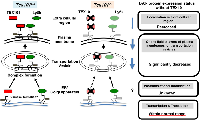
Figure 5. Molecular status of Ly6k with/without TEX101 in the TGCs.
The left diagram indicates TEX101/Ly6k complex formation of wild-type (Tex101+/+) mouse. After translation, GPI remodeling of these molecules is completed from ER to Golgi apparatus, then these molecules are expressed as TEX101/Ly6k complex (represented by black square) on lipid bilayers including transportation vesicle and plasma membrane. In addition, (a part of) both TEX101 and Ly6k are released into extracellular space. In TEX101-null TGCs (the right diagram), Ly6k expression is drastically decreased. Black cross marks indicate the disruption of the molecules. The potential status of Ly6k protein expression without TEX101 is boxed.

