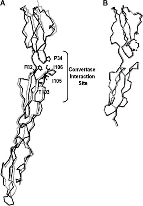
Fig. 1.
Homology model of N-terminal domain of CR1. (A) C-α tracing of SCR-1–SCR-3 domains of CR1 (black) superimposed on the X-ray crystal structure (grey) of human complement regulator CD55. The residues, forming the hydrophobic patch and responsible for interaction with convertase, are shown as ball-and-stick model. (B) C-α tracing of SCR-1 and SCR-2 domains of CR1 (black) superimposed on the NMR structure (grey) of SCRs-15–16 of CR1.

