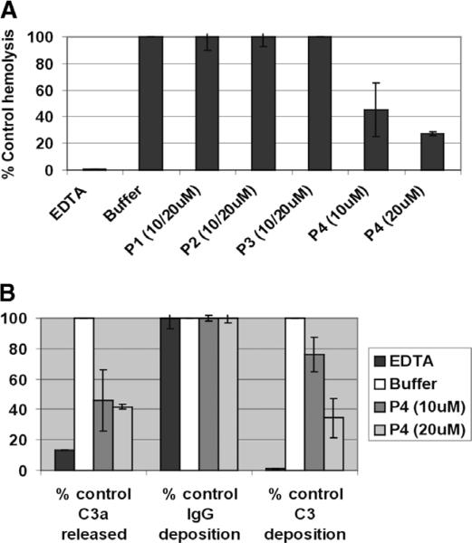
Fig. 3.
Anti-complement activity of CR1-derived peptides. (A) Human sera from a group O donors (=2) were pre-incubated for 15 min at room temperature with control 10 mM EDTA (for inhibition of the complement activation pathways), 10 or 20 μM peptides 1–4, before addition of human group A RBCs. Levels of hemolysis in the supernatants were measured. Data are presented as the percentage of hemolysis the buffer control from four separate experiments. (B) Surface C3 and IgG were measured on the remaining unlysed cells from P4-treated samples in A by flow cytometry. Levels of C3a released in the supernatants were measured using an ELISA kit. Data are presented as the percentage of C3 and IgG deposition and C3a release of the buffer control from four separate experiments.

