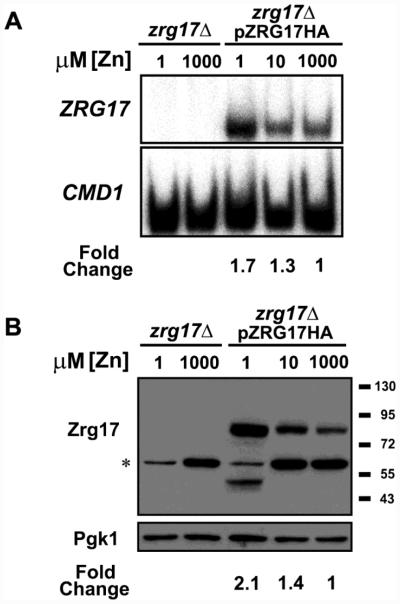
Figure 2. Zrg17 protein levels correlate with ZRG17 mRNA.
zrg17Δ mutant cells or zrg17Δ cells expressing a C-terminal hemaggluttinin (HA) epitope-tagged allele of Zrg17 expressed from its own promoter (pZRG17-HA) were grown in LZM supplemented with the indicated concentration of ZnCl2. A) S1 nuclease protection assays were performed to detect ZRG17 and CMD1 mRNA. The fold changes shown were quantified from the level of ZRG17 mRNA normalized to CMD1. B) Immunoblot analysis of Zrg17-HA protein levels. Lysates were prepared from the same cells used in panel A and subjected to immunoblotting with anti-HA antibody. Pgk1 (phosphoglycerate kinase 1) served as a loading control. The asterisk indicates a non-specific background band. The fold changes shown were quantified from the level of Zrg17 protein normalized to Pgk1. A smaller molecular mass Zrg17-HA band was observed in the LZM + 1 μM ZnCl2-grown pZRG17-HA transformants, perhaps resulting from partial proteolysis, and both full-length and truncated Zrg17 forms were included in the quantification. The data shown are representative of two independent experiments.

