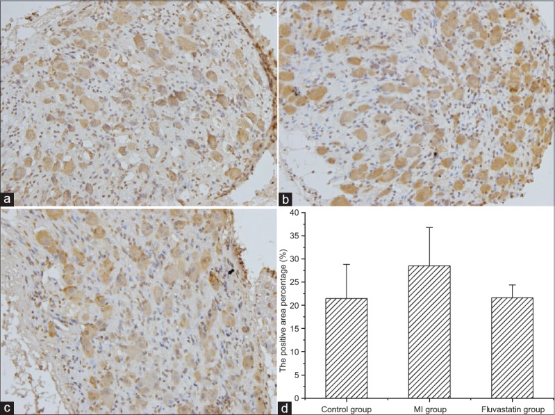
Figure 4.

Immunohistochemical staining of Nav1.7 protein in stellate ganglion (original magnification, ×400). Typical examples of Nav1.7 protein in control group (a), myocardial ischemia group (b), and fluvastatin group (c). (d) Histogram of the percentages of Nav1.7 protein in three groups. The percentages of Nav1.7 protein in control group, myocardial ischemia group, and fluvastatin group were 21.49 ± 7.33%, 28.53 ± 8.26%, and 21.64 ± 2.78%, respectively (n = 5 in each group, F = 1.495, P = 0.275).
