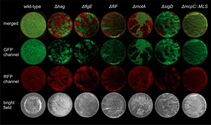
Fig. 5.
Stereomicroscopy images of B. subtilis wild-type or mutant strains competed against its genetically identical, but differently labelled derivative. Wells (16 mm diameter) of a 24-well plate are shown in the green- or red-fluorescence channels (false-coloured in green and red) together with merged and bright light images. Representative examples for each strain are shown.

