
Figure 14.
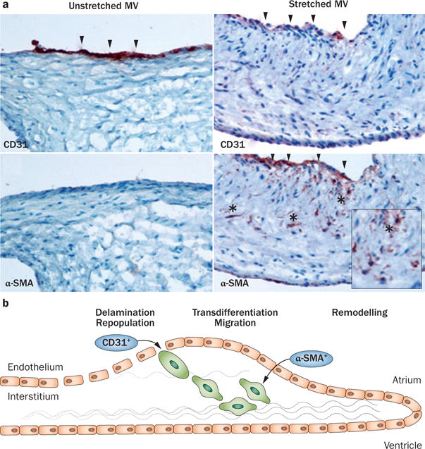
Evidence for reactivated EMT owing to leaflet stretch. a | Staining for the endothelial marker CD31 is reduced in the atrialis, and staining for smooth muscle α-actin is detected, which is normally absent, with positive cells (*) migrating into the interstitium, all features of EMT. Left panels: unstretched MV showing no staining for α-SMA along the CD31+ endothelium. Right panels: staining for α-SMA in the atrial endothelium (also CD31+) of a stretched MV, with nests of cells positive for α-SMA seeming to penetrate the interstitium (*). Upper panels: arrowheads indicate CD31+ endothelial cells. Lower panels: arrowheads indicate α-SMA+ endothelial cells, indicative of EMT. b | Schematic of active MV adaptation by EMT. Abbreviations: α-SMA, smooth muscle α-actin; EMT, endothelial-to-mesenchymal transition; MV, mitral valve. Reprinted from Dal-Bianco, J. P. et al. Active adaptation of the tethered mitral valve: insights into a compensatory mechanism for functional mitral regurgitation. Circulation 120 (4), 334–342 (2009). Bottom panel modified from Armstrong, E. J. & Bischoff, J. Heart valve development: endothelial cell signaling and differentiation. Circ. Res. 95 (5), 459–470 (2004).