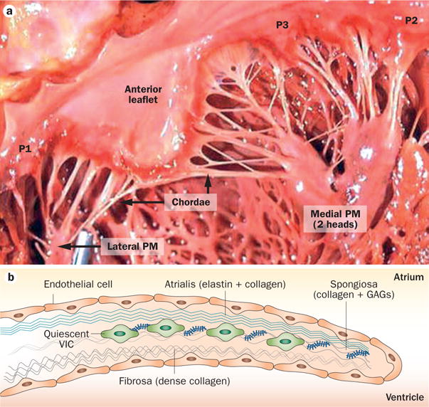
Figure 2.

Mitral valve structure. a | The mitral valve has two leaflets—the anterior leaflet below the aortic valve, and the posterior leaflet composed of three scallops: P1, P2, and P3. At the leaflet edges, the chordae tendineae link the leaflets to the PM anchors on the left ventricular wall. b | The leaflet structure is well organized, with a dense fibrous layer of collagen, known as the fibrosa, which arises from the mitral annulus and faces the left ventricle. The fibrosa is thicker close to the annulus and thinner at the edge of the leaflet. The fibrosa is covered by the spongiosa, a loose connective tissue layer rich in GAGs. The spongiosa is thicker at the leaflet edge and thinner at the annulus. The atrialis is made up of lamellar collagen and elastin sheets, which extend from the left atrial endocardium into the leaflet. Endothelial cells cover the valve. The deep subendothelial layers contain quiescent VICs—noncontractile, fibroblast-like cells that originate from endocardial endothelial cells. Abbreviations: GAG, glycosaminoglycan; PM, papillary muscle; VIC, valvular interstitial cell.
