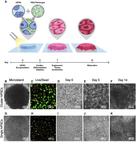
Fig. 1.
Schematic and progression of hiPSC encapsulation to produce 3D-dhECTs. (A) HiPSCs were combined with aqueous PEG-fibrinogen precursor, added into a PDMS mold (not shown) on acrylated glass, and photocrosslinked using a non-toxic photoinitiator and visible light to form 200 μm thick “microisland” tissues. All encapsulated hiPSCs were maintained in their pluripotent state for three days, followed by initiation of cardiac differentiation to produce uniformly contracting 3D-dhECTs. Encapsulated (B) cluster and (G) single hiPSCs formed “microisland” tissues (images taken on day 0 of differentiation). (C, H) HiPSCs remained viable (green) 24 h post-encapsulation (n = 3 tissues) and (D-F, I-K) proliferated during early stages of cardiac differentiation. F, K represent Movies S3 and S4.

