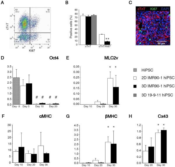
Fig. 2.
3D-dhECTs enabled efficient cardiac differentiation and gene expression. (A) Representative flow cytometry results from day 20 3D-dhECTs. (B) 3D-dhECTs and aged-matched 2D monolayers (control) showed comparable differentiation efficiency on day 20 of differentiation. n = 3-5 biological replicates per group. Mean ± s.d. ANOVA P < 0.05, ** vs. 2D (C) Cardiac marker cTnT and proliferation marker Ki67 were also observed using immunofluorescence staining of day 20 cardiac tissues. (D) Pluripotency gene Oct4 decreased following initiation of differentiation, while cardiac genes (E) MLC2v, (F) αMHC, (G) βMHC, as well as (H) the functional gene Cx43 showed trends towards CM maturation over time. All mRNA levels were normalized to the housekeeping gene GAPDH. n = 3 biological replicates per group. Mean ± s.d. ANOVA P < 0.05, # vs hiPSCs and day 0; * vs. earlier time point.

